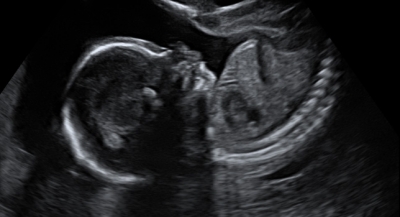
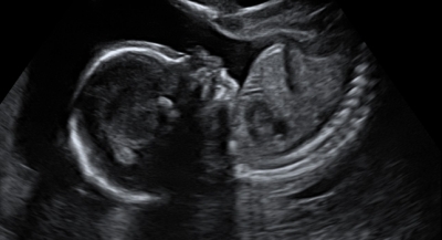
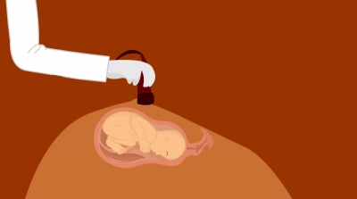
At the end of the first trimester during 11 to 13 weeks, a screening test is done which includes an ultrasound looking for nuchal translucency(it is a collection of fluid under the skin of the baby’s neck). All developing babies have some fluid at the back of their neck. But many babies with Down's syndrome have an increased amount of fluid and can lead to an increased nuchal translucency as seen in the ultrasound. NT scan is usually done around 11 to 13 weeks of pregnancy. After 14 weeks the scan may not be reliable to detect Down’s syndrome.
The first-trimester screening also includes a blood test which measures the levels of HCG, AFP and PAPP- A. This is done to screen for any chromosomal abnormalities or heart defects. Thus the first-trimester screen includes the ultrasound test and a blood test done during the 11th to 13th week.
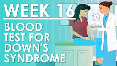
The second-trimester triple screen or quad screen is done during the 16th to 18th week. This screening includes a blood test which measures the levels of AFP, HCG, Estriol (predominant estrogen during pregnancy) and inhibin A. Some doctors do both the first trimester and second-trimester quad screen. There is conflicting evidence whether one is more accurate than the other.
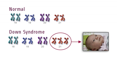
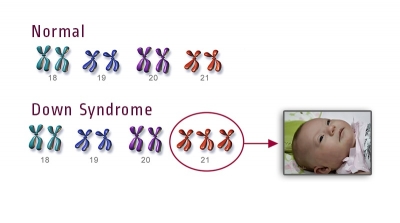
During the Second trimester usually ,two tests are done - one is a quad screen or triple screen(to detect aneuploidy) and the other is an ultrasound which is called fetal anatomy ultrasound done at the last part of the second trimester. In the quad screen - AFP is most imp of all. High or low levels of AFP can indicate an increased risk for certain conditions in the baby. AFP is a Serum glycoprotein produced by the embryo. It gets to a peak concentration in the amniotic fluid by 12 weeks and it peaks in the maternal serum by about 30 weeks. AFP levels continue to increase during the whole of the second trimester and thus dating of pregnancy is very important, as AFP levels change with time and what is normal for one time in the duration of pregnancy may be high or low for another week of pregnancy. Inaccurate dating of pregnancy is one of the most important causes of abnormal levels of AFP. AFP levels are calculated as median AFP levels. If it is 1 it is normal, if it is significantly lower than 0.85 multiples of the median, it could be that the pregnancy is at increased risk for ( Down’s syndrome, inaccurate gestational age, Turners syndrome). When the Doctor finds out the levels of Median AFP levels are lower than 0.85 median AFP levels then usual next step is to do a sonogram to confirm the gestational age - Sonography can also look for Nuchal translucency and nuchal fold. If sonogram confirms gestational age, and if the levels are low, then the mother may be advised to go for amniocentesis to get the karyotyping, which may be diagnostic for Downs syndrome or Turners syndrome. Down’s syndrome will have an extra 21st chromosome
If the AFP levels are more than two half times the median (more than 2.5 ) then it is considered high and reasons for this could be inaccurate gestation, multiple gestations, neural tube defects, ventral wall defect, pataus syndrome and fetal renal diseases, placental bleeding. Next step, in this case, is again to go for sonography to confirm the gestational age and also look for multiple gestations or anomalies. If sonography is normal then an amniocentesis is done to look for levels of Amniotic fluid AFP levels and an enzyme called Acetylcholine esterase. High levels of these in the amniotic fluid will be diagnostic of Neural tube defects and if the levels are normal it could be a normal pregnancy, but this pregnancy may carry an increased risk of IUGR (intrauterine growth retardation), SGA(Small for Gestation), Stillbirth and pre-eclampsia. Why does it increase in Pre-eclampsia - probably because AFP is a glycoprotein made by the placenta and we know that in Pre-eclampsia there is a problem with the placenta
Both in Down’s syndrome and Turners syndrome show a similar profile on the quad screen - i.e the levels of estriol and AFP are low and the levels of HCG and inhibit are high. The sonogram also may look alike, that is why it is very important to do an amniocentesis to determine the karyotype. Downs syndrome is however more common than Turners syndrome. In Edwards syndrome Estriol and HCG levels are low and in Patauss syndrome except for AFP levels all the other three levels are normal. AFP levels are high.
Second-trimester ultrasound is performed towards the end of the second trimester at around 18 to 22 weeks. This ultrasound is part of a routine antenatal care. One looks at the fetal cardiac activity, fetal number, anatomy of fetal organs, fetal biometrics ( HC, BPD, FL Abdominal circumference) to estimate the fetal weight, placental appearance location, amniotic fluid, uterine anatomy and lower segment etc
Most Pregnant women are offered both the first trimester and second-trimester screening tests. It is important to consult your doctor and decide which is right for you.

