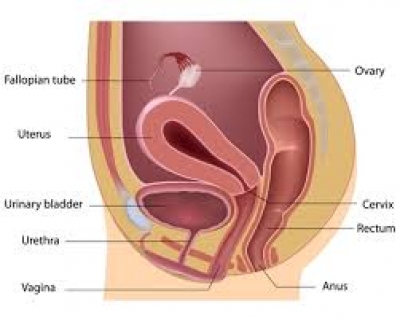
It may also get implanted on the ovaries, outside the uterus. The doctor will also look for cardiac pulsations during this time.
Thus the location of the pregnancy is very important. The gestational age of the pregnancy is calculated by measuring the crown-rump length(CRL). This measures the distance between the top of the head of the baby to the lower end of the sacrum. From 6 to 11 weeks gestational age, the fetal CRL grows at a rate of about 1 mm per day. So usually the gestational age is calculated by adding the CRL to the 6 weeks basic age. For eg - if the CRL is 25 mm then the gestational age will be 9 weeks 4 days ( 6 weeks + 25 ( 3 weeks(21 days) +4). After 12 weeks the Biparietal diameter is used to measure the gestational age (measurement of the width of the fetal head).
The Nuchal scan is sometimes done during the 11 to 13 weeks and 5 days of pregnancy. The timing is very important for this scan as the doctor will look for certain things which are only seen during this time period. This test is usually done for screening for Down’s syndrome.
A collection of fluid beneath the fetal skin in the region of the fetal neck is called Nuchal Translucency. This is present in all fetuses in early pregnancy. But the increased collection of fluid in this region could be an indication of Down’s syndrome or other chromosomal abnormalities. It appears in the ultrasound as a black space beneath the fetal skin. The black space will be measured during the ultrasound scan. This is normally less than 2.5mm and when seen increased (greater than 2.5mm) may indicate the baby has Down syndrome or may indicate another chromosomal abnormality. Currently, this is probably the most accurate noninvasive test for detecting Down syndrome during pregnancy. They also check for visible fetal nasal bones during the Nuchal scan. Presence of fetal nasal bones decreases the chances of Down’s syndrome.
The nuchal translucency test can also check whether the baby has all its limbs, and also see if the head and brain appear to be developing normally, that the baby has a visible stomach and bladder and the umbilical cord is inserted correctly. They also check the spine to rule out spina bifida.
Along with the NT scan, the doctor may also recommend a blood test to test for the levels of two hormones released by the body during pregnancy. These are namely PAPPA (Pregnancy-Associated Plasma Protein–A) and free beta–hCG ( Human Chorionic gonadotropin) The two hormones measured in the blood are both produced by the placenta and are unique to each pregnancy. If the two hormone levels are favorable they will reduce the chance of Down's syndrome. If they are less favorable they will increase the chance. Adding in the blood test to the NT scan increases the overall accuracy of the test.
[quix id='8' name='About our Pregnancy Blogs and Videos']


